| Quick overview of presentations given by IFMP faculty |
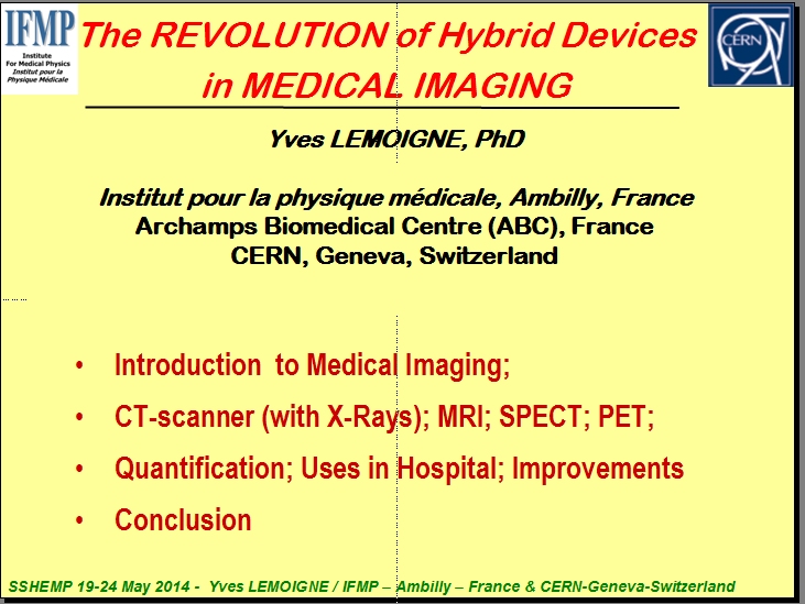
First Page of Power-point talk |
Photo |
Short text |
|
 Yves
Lemoigne Yves
Lemoigne
IFMP Ambilly
|
This is an introduction to the Medical Physics program.
The teacher presents a review of different imaging systems and modalities (e.g., X-ray computed tomography (CT), Magnetic
Resonance Imaging (MRI), Single Photon Emission Tomography (SPECT) and Positron Emission Tomography (PET)), their operating
principles, their use in the hospital (diagnosis, treatment -planning, post- treatment survey), while also emphasizing their complementarities: X-ray computed tomography (CT), Magnetic Resonance Imaging (MRI), Single Photon Emission Tomography (SPECT) and Positron Emission Tomography (PET). The significant progresses and innovations in this area during the last decade are further discussed: Photon time of flight measurement, hybrid combination of complementary devices like SPECT-CT, PET-CT and PET-MR, etc).
Many of these points will be developed separately by the following speakers. |
| For whole talk: |
click here
|
 |
 Yannick Arnoud Yannick Arnoud
LPSC, CNRS, Grenoble |
The teacher explains the basics on Positron Emission Tomography (PET) :
- Metabolism imaging (Role of Glucose in Need for energy from Any active tumor)
- Radiotracers (Including FDG and recalling operating principles of PET)
- Sinogram (for image reconstruction)
- Correcting for detector effects
- Attenuation map
- Quantification
- Why PET is a nuclear medicine precious tools. |
| For whole talk: |
click here
|
 |
 Alex Rijnders Alex Rijnders
Europe Hosp. Brussels |
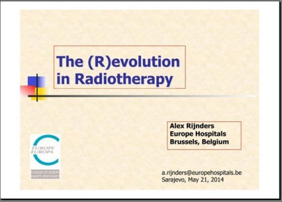
The teacher first recalls the goals and backgrounds of Radiotherapy(RT). He explains the conformal radiotherapy (3D - CRT)
whose principle is: to conform with a high precision the spatial distribution of the prescribed dose delivered to the 3-D target
volume, while excluding critical structures as far as possible. (Possibility to improve local control and outcome).
Only technological improvements allowed its technical realization: precise imaging, fast computers for treatment planning,
Multi-Leaf Collimators (MLC), Electronic Portal Imaging (EPID) ...
Intensity Modulated Radiotherapy (IMRT) was the next progress, i-e a special form of conformal therapy using geometric field
shaping + intensity variation modulation. All that helps to overcome some limitations of 3D-CRT. Intensity of the primary photon
fluence varies across the target volume (each beam may treat only portions of the target). Several examples are given.
Then Image-Guided Radiotherapy principles are explained (IGRT), correcting for patient positioning errors. This allows reduction
of safety margins and better probability to hit the target. Combination IGRT + IMRT = High Precision RT (Lower probability of side
effects, Higher dose to tumor).
|
| For whole talk: |
click here
|
 |
 Yves Lemoigne
IFMP, Ambilly Yves Lemoigne
IFMP, Ambilly |
Biomedical research needs a precise quantification of biological parameters and PET can provide them.
As example, the speaker refers to an experience of neurotransmission in the brain of a rat. The aim was to study the binding of
serotonin, in fact quantifying 5-HT1A receptors interactions in rats. Serotonin is involved in various physiological functions
and behaviour like anxiety, depression (to fight against)
This will :
1- Rely on backgrounds and mathematical modeling (basics of pharmaco-kinetics); establish the need for dynamic imaging (i-e change
in a given period of time); role of mathematical models.
2- Discuss the principles of the experiment and data analysis from micro-PET: measurement of the density of 5-HT1A receptors expressed
in pico-mol by ml in the hippocampus area of a rat brain. |
| For whole talk: |
click here |
 |
 Alex Rijnders Alex Rijnders
Europe Hosp. Brussels
|
Review of current / futuredevelopments in BrachyTherapy (BT):
Sources - Isotopes (classical and new isotopes, electronic BT sources, seeds ...)
Afterloaders / applications (HDR, PDR ...)
Use of modern imaging technologies / tools: Image-guided Brachytherapy (X-ray, CT spiral, multislice, with 0.5T MR open, US, PET).
Robotics?
- Dose Calculation (GST): 3D prostate treatment planning BT, BT dose calculation: TG43
-Training in Brachytherapy: with technical treatment and delivery systems becoming more complex, there is huge need for better
formed / trained staff (few specialised centers in Western Europe). |
| For whole talk: |
click here |
 |
 Sonja
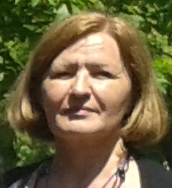
Petkovska Sonja
Petkovska
ACIBADEM Skopje |
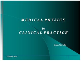
Experience as Medical Physicist in clinical practice. The importance of
physics / physicists in clinical practice is stressed, especially
increases necessity of medical physicists for using radiation in:
- diagnoses
- treatment
* Protection from radiation (patients, workers, public)
* In diagnosis (2D radiographic, CT, MRI, PET): adjust the parameters
- Devices: commissioning, calibration, periodical measurements
- Survey: patient position, patient dose (as less as possible)
* In treatment (simulation, verification, evaluation)
- Pretreatment imaging for planning (CT, PET, MRI)
- Examples
- Benefits of Combined Technique
* Imaging CBCT for treatment verification |
| For whole talk: |
click here |
 |
 Alejandro
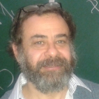
Mazal Alejandro
Mazal
Curie Inst. Paris |
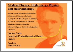
HadronTherapy (HT): The goal of the present lecturer is not to teach “hadrontherapy” not thoroughly in one hour
but to give as a “taster - fast movie” of this fascinating and powerful approach to RT and give the wish to know more!
1. Medical Physics in RT : past, present
- The technological evolution of external radiation therapy 100 years of history
Ii) Particles types: Pions, Neutrons, Protons, Lights & Heavy Ions.
ii) Interactions with electric (E) and magnetic fields (B) / Interaction with matter (e-g: beam line, patient)/ Cyclotrons / Synchrotrons
/
2. Hadrontherapy:
A typical hospital-based protontherapy centre (Loma Linda, USA): i) Dose distribution in nanometre scale
ii) Bragg Peak (Characteristics, Spread-out, Beam-Target interactions, Spot..)
/
Intensity Modulated IMPT-IMZT /
Planning and treating with particles: examples of clinical applications /
Moving towards heavier ions
3. The future revisiting radiation biology /
Relative Biological Effectiveness (RBE) Particles: LET dependencies /
Use of nanoparticles to improve the performance of hadrontherapy /
between basic research and medical physics with engineering, /
biologists and physicians, institutional and political administrators
|
| For whole talk: |
click here |
** Quick overview of the presentations by students participating in Working Groups |
 |
 Hoda
AbouShady Hoda
AbouShady
Cairo Univ. |
Prof. Hoda Habou-Shady has presented the Center of Nuclear Studies and Peaceful Applications (NSPA) in Cairo University.
She is the founding-director of this Center.
The mission of the centre is to establish a research facility for civil applications in nuclear sciences. This centre will be the
first center in academia that supports training on medical physics, radiation protection, dosimetry and bioethics. It will also include
simulation and computational techniques needed in the training of physicist in various fields of nuclear applications. The centre also
includes other applications such as in synchrotrons and accelerators and material sciences.
The centre is organised in 6 departments: 1- Nuclear Science Department 2- Nuclear Technology Department
3- Research and Projects Department 4- Training and Consulting Department 5- Radiation Protection and Dosimetry Department
6- Environment and Sustainable Development Department
To see Prof. Hoda interview about Nuclear Energy at Nile TV :http://www.youtube.com/watch?v=AQcoKIFusNA&feature=channel&list=UL |
| For whole talk: |
http://www.youtube.com/watch?v=AQcoKIFusNA&feature=channel&list=UL |
 |
 Philip Puicea Philip Puicea
IFIN-HH Bucaresti |
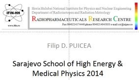
Philip Puicea presents a Romanian Radiofarmaceuticals Research Center. It will include a cyclotron where Particle acceleration
is achieved using an RF field between “dees” with a constant magnetic field to guide the particles. He shows the TR-19
Cyclotron which has been chosen: 1- Fully automated and computer controlled machine.
2- Variable Energy: extraction energy between 14 MeV and 19 MeV.
3- External Ion Source (H-) and Extraction System. 4- Automated Target Changer with precise real-time target position alignment.
5- Gaseous and liquid targets (solid targets processing system). 6- Experimental hall with additional beam lines.
Schematic of Drug Structure for PET Imaging: Molecular Address => Reporting Unit
It will include a chemical lab with automated synthesis Modules and an automated dispensing unit for delivering of radiopharmaceuticals.
|
| For whole talk: |
click here |
 |
 Bistra Hristova Bistra Hristova
PHRH- Varna
|
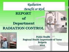
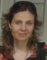
Bistra Hristova explains the Department of Radiation Control from Regional Health Inspectorate of Varna, Bulgaria. She is in charge of
the department.
The major task of the Department of Radiation Control is to protect and promote the physical and environmental health of the people.
There are two groups of Radiation control:
-1- First group in charge of:
- Control before the execution of a project
- Current Radiation Control
- Radiation control in general (industry, science, medicine, agriculture)
- Comprehensive Radiation Control (beta equipment for analysis)
|
| For whole talk: |
click here |
 |
 Marina
Zdraveska Marina
Zdraveska
Skopje Univ. |
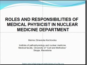
Marina Zdraveska Kochovska presented the Department of Medical faculty, University of "Cyril and Methodius" in Skopje,
Macedonia. She reviewed the staff and equipment of her service: Four gamma cameras, one Mediso Planar, Two SPECT
(Sopha DS7 and SIEMENS ecam) and one dual head SPECT (Mediso DHV). She detailed the task of a Medical Physicist in the Nuclear
Medicine Service with examples from Tc99m MIBI Myocardial perfusion but also examples related to Su COLOID Tc 99m liver or
Tc99m DTPA Renal studies, the latter being achieved by dynamic acquisition.
She detailed the charges of a medical physicist:
- Dosimetry control and monitoring (patients, workers and environment)
- Quality control of equipment
- Radiation protection and safety
- Radioactive waste
- Research and education< |
| For whole talk: |
click here |
 |
 Nikolina
Sarcevic Nikolina
Sarcevic
Novi Sad Univ |
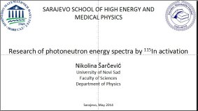
Nikolina Sarcevic presented a report about the research of photoneutron energy spectra by In-115 activation. Questions:
Neutrons / Medical linear accelerators (linacs) /
Photo-neutron spectra comparison of two different methods
She Explained linacs 101 focalizing on Linac Head: 1- Absorbers (Al, C)
2- Collimators (primary, jaws, MLC) 3- Flattening filter (etc.)
4- Detector (ionizing chamber) 5- Target (X-ray or electron mode)
Principle: High Energy Phot => High Z material =>Photoneutrons
Motivation: - Neutron quality factor is 20
- Safety issues (staff at maze door)
- Secondary cancers
What she recommended:- Re-test / calibrate Bonner sphere gold foil activation technology
- Measure neutron spectra 1m SAD / 40 cm off isocenter using In-115 activation
- Recheck Before publishing data (!) |
| For whole talk: |
click here |
 |
 Lejla Civa Lejla Civa
Sarajevo Univ |
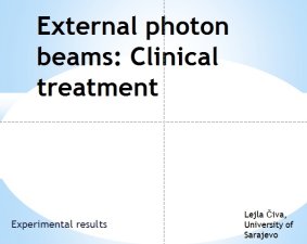
Lejla Civa presents a study on external photon beams in a clinical treatment.
Photon sources are either isotropic or non-isotropic and they emit either monoenergetic or heterogeneous photon beams.
In external beam radiotherapy, photon sources are often assumed to be point sources and the beams they produce are
divergent beams. The photon fluence is inversely proportional to the square of the distance from the source.
The surface dose represents contributions to the dose from:
● Photons scattered from the collimators, flattening filter and air;
● Photons backscattered from the patient;
● High energy electrons produced by photon interactions in air and any shielding structures in the vicinity of the patient.
In Radiotherapy, a percentage depth dose curve (PDD) (sometimes percent
depth dose curve) relates the absorbed dose deposited by a radiation beam
into a medium as it varies with depth along the axis of the beam. |
| For whole talk: |
click here |
 |
Mahira Tvirtkovic
Sarajevo Univ
|
 |
For whole talk |
|
|

 Yves
Lemoigne
 Yannick Arnoud

 Alex Rijnders

 Yves Lemoigne
IFMP, Ambilly
 Alex Rijnders
 Sonja
Petkovska
 Alejandro
Mazal
 Hoda
AbouShady

 Philip Puicea

 Bistra Hristova
 Marina
Zdraveska

 Nikolina
Sarcevic
 Lejla Civa

