** Quick overview of presentations given by IFMP faculty |
First Page of Power-point talk |
Photo |
Short text |
 |
Y-Lemoigne |
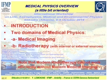
The lecturer introduces the Medical Physics program. He presents a review of Medical Imaging which will be the main topic of this Medical Physics part of the workshop.
Five different techniques will be studied: Ultrasound, CT, SPECT, PET, MRI. Then we will see their use in Hospital.
IMAGING Exploration inside the human body is done within two purposes:
1- ANATOMICAL to see INSIDE the body at a moment (X-rays CT, MRI, Ultrasounds)
2- FUNCTIONAL to see how the body functions during a period of time (PET, SPECT, fMRI.)
There are two types of machines depending if source is OUTSIDE the body like the
detectors (CT for instance) or INSIDE the body (PET or SPECT).
Ultrasound will be explained in details by Christian Cachard and Hervé Liebgott.
Ultrasound are are popular because 3 POSITIVE POINTS which are:
-
Real time imaging;
- No radiation
- Relatively low cost (no special building requested).
APPLICATIONS at HOSPITAL or Private CLINIC: Two talks will be given by D. Miladinova and S. Petkovska on Wednesday. They are both from Skopje, Macedonia.
Note that a specific talk about MRI
will be given by Kyriaki Theodorou on Tuesday.
INNOVATION in THERAPY: with Ultrasons (HL), with Hadrontherapy (PA, KT)
|
| For whole talk: |
click here |
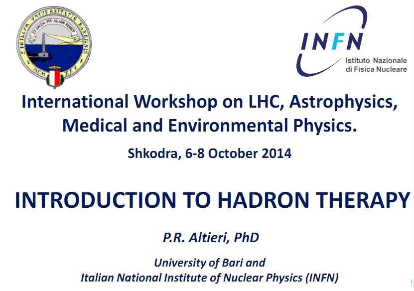 |
Palma Altieri
|
Hadron therapy is increasingly considered the best radiotherapy for cancer due to its superior dose distribution. Compared to photons or electrons used in the conventional radiation therapy, the hadrons (mainly protons, neutrons and light ions) have a depth dose profile such that the energy is released to the tumor target with a high accuracy, so avoiding the surrounding healthy tissues.
The aim of this talk is to give an overview of the hadron therapy starting from the history of such a therapeutic treatment. Afterwards the physical basics will be described as well as the biological properties, in order to show the advantages of protons and carbon ions with respect to X-rays in radiation oncology.
Finally the most interesting results achieved in this multidisciplinary research field will be discussed and the future challenges introduced.
|
| For whole talk:
|
click here
|
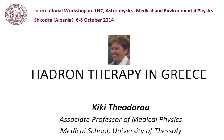 |

K-Theororou |
Hadron therapy is the most advanced
contribution of technology and innovation from the field of experimental
particle physics to the fight against cancer, explaining the increased
interest for the development of such installations worldwide.
The presentation will be focused on cancer epidemiology in Europe and in Greece, on the radiobiological advantages provided by hadron irradiation, on the feasibility study and the requirements for the installation of a hadron therapy facility. |
| For whole talk: |
click here
|
 |
Christan Cachard
|
The place of ultrasound in medical imaging is important because:
* Ultrasound has the last decade been the fastest growing imaging modality for non-invasive medical diagnosis.
* Of all the various kinds of diagnostic produced in the world, one of four is an ultrasound scan.
* Reasons for this are the ability to image soft tissue and blood flow
* the real time imaging capabilities,
* the harmlessness for the patient and the physician (no radiation)
* the low cost of the equipment.
* no special building requirements as for X-ray, Nuclear, and Magnetic Resonance imaging.
* Limitations are that ultrasound imaging cannot be done through bone or air (limitations on chest imaging).
* Ultrasound imaging depends on acoustic wave backscattered toward the probe by scatterers
*Increasing the amount of scatterers should increase the number of echoes and therefore, improve the image.
|
| For whole talk: |
click here
|
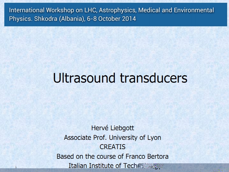 |
Hervé Liebgott |
This lecture is about Transducers in Ultrasound: Probe types and anatomy
. Transducer models and Structure (finite elements)
- Impulse response (acoustics)
- Beam profile (aperture)
- Transducer structure and characterization and Technological aspects
- There is a multitude of available probes, but they all share some common properties:
i° They are made of
piezoelectric material ; ii) They comprise many active elements; iii)
various devices for impedance matching and
focalisation are present; iiii)Safety and ergonomic aspects regulate the physical characteristics of the probe
|
| For whole talk: |
click here
|
|
Christan Cachard |
Lecture on Basis of ultrasound and medical ultrasound imaging (see previous Cachard talk)
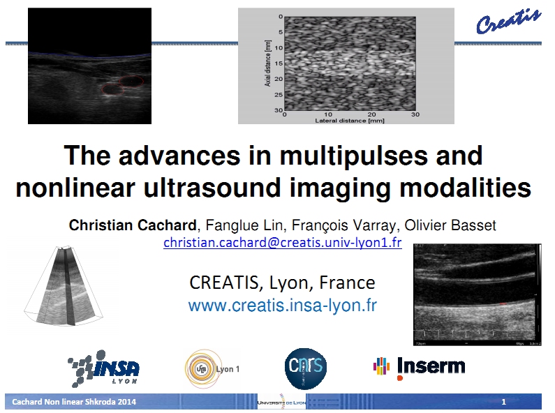
* Advanced in Ultrasound: Contrast agents are useful in all other imaging modalities (X ray, MRI, PET, ...)
* Ultrasound Contrast Agent :Increasing the amount of (strong) scatterers should increase the umber of
echoes and therefore, improve the image.
- Nonlinear ultrasound
* Improvement of ultrasound imaging or Nonlinear imaging: i) Harmonic imaging;
ii)Multipulses imaging or
Contrast (nonlinear) imaging; iii) Pulse inversion (Pl); iv) Second Harmonic Inversion (SHI):
v) Amplitude
modulation (AM); vi) Pulse inversion amplitude modulation (PIAM); vii) Phase coded sequences (PCS);
contrast
pulse sequence (CPS)
* Multipulses imaging or Contrast (nonlinear) imaging
* Generalization of multi-pulse techniques
* Influence of scatterer motion to phased multipulses method
|
| For whole talk: |
click here
|
  |
Hervé Liebgott |
This lecture is about examples and the corresponding evolution of ultrasound imaging
*Static elastography:Limitation of static elastography: i)Only qualitative
/ relative stiffness; ii) Depends on
the applied load; iii) Not quantitative
- *Shear wave elastography: The idea of shear wave elastography; Shear wave
propagation velocity is prop
portional to shear modulus
. *Tissue elasticity imaging - clinical motivation- The objective of elastography is to produce a map of the stiffness of tissues- There is strong correlation between stifness and some pathologies
. * *ultrafast imaging; photo-acoustic imaging: Ultrafast imaging can do many things (2 examples):
i) Complex
motion visualization of Blood; Cardio ; Arterial wall motion; ii)
Functional imaging of the Brain / Activation of the different parts of the brain is linked with an increase in blood flow;/Power
Doppler can give an indication of the quantity of flow in a region;
- *Functional imaging of the brain with ultrasound is doone by combining ultrafast imaging and power Doppler. Example in the case of micro vascularization of trepanned rat brain during whisker stimulation
|
| For whole talk: |
click here
Lecture about Ultrasonic Doppler:
.i) What is Doppler effect. ii) CW/PW Doppler systems building-blocks.
iii) Pulsed Wave (PW) mode:
-PRF, sample volume, spectral broadening, mean frequency estimation...
- Advanced Doppler systems and methods:
~ Single-gate (TCD, Duplex),
~ Multi-gate,
~ Flow-imaging,
~ Power, Harmonic & Tissue Doppler imaging.
~ Doppler artefacts (aliasing, blooming...) |
|
Christan Cachard
|
|
| For whole talk: |
click here
|
 |
Hervé Liebgott |
Transducers: Probe types and anatomy
~ Transducer models
- Structure (finite elements)
- Impulse response (acoustics)
- Beam profile (aperture)
- Transducer structure and characterization
- Technological aspects
~ There is a multitude of available probes, but they all share some common properties:
- They are made of piezoelectric material
- They comprise many active elements
- Various devices for impedance matching and focalisation are present
~ Safety and ergonomic aspects regulate the physical characteristics of the probe
|
| For whole talk : |
click here
|
 |
K-Theororou |
Magnetic Resonance Imaging has gradually became a valuable tool for diagnosis. The recent years, through of the availability of continuously higher magnetic fields, more qualitative and quantitative imaging techniques have been developed.
Today, the clinical availability of more than 3T fields enable scientists to perform advanced MRI techniques, i.e. MRS, DTI, DWI etc, thus contributing to differential diagnosis.
This presentation will focused on the application of those advanced techniques and especially to the differential diagnosis of the central nervous cases. |
| For whole talk |
click here
|
 |
D-Miladinova |
UNIVERSITY Ss CYRIL and METHODIUS, Skopje, Macedonia
. APPLICATIVE PHYSICS IN CLINICAL PRACTICE (in nuclear medicine)
- Radioactivity (physical characteristics, radioactive decay, interaction of radioactivity with matters, types of detectors, collimators, counters, spectrometry, statistics).
- Scintigraphic systems (scanners, gama cameras, counters, PET camera, physical parameters of scintigraphic systems, quality control in scintigraphic systems,systems for image presentation.
- Computers in nuclear medicine (computer analysis of acquired data, static and dynamic data, filters for processinfg, improvement of image quality, deconolution analysis, amplitude, phase analysis, methodt for simulation of biological systems, acquisition and data analysis in PET).
. APPLICATIVE PHYSICS IN CLINICAL PRACTICE (in radiotherapy)
- Radionuclides in radiotherapy: 2 months
- Dosimetry of after-loaded sources: 4 months
- Dosimetric control of radioactive sources: 1 months
- Equipment for percutaneous radiotherapy: 4 months
- Dosimetry of percutaneous radiotherapy: 4 months
- Diagnostic radiology: 4 months
- Computers in radiotherapy: 2 months
. UNIVERSITY CLINIC of RADIOTHERAPY & ONCOLOGY
with 4 specialists, 1 on specialization, 5 physicists
. INSTITUTE of PUBLIC HEALTH: Laboratory for radioecology; Laboratory for ionizing radiation |
| For whole talk |
click here
|
 |

Sonja Petkovskac |
. To be a Medical Physicist you should:
- Have a high level of ability and interest in physical sciences and computing
- Have medicine interest & development of new methods of patient care and treatment
- Be accurate, able to concentrate for long periods, have a high level of attention to detail
. What does it take to become a Medical Physicist?
- Have high ethical standards and the ability to take responsibility for making decisions
- Have an enquiring mind and good problem solving skills
- To lead a research and development team
- Have excellent oral and written communication skills
- Be able to reassure nervous patients.
. What does a Medical Physicist do?
- Apply the principles of physics to patient care
- Work closely with doctors and other professionals to assess and treat ill health research.
- Design, develop technical equipments used by medical staff to diagnose and treat patients
(alongside other specialist physicists, clinical engineers and technical staff) |
| For whole talk: |
click here
|
 |
Y-Lemoigne |
Shkodra workshop focussed on Medical Imaging and innovative Radiotherapy.
in Medical Imaging we preferred the ultrasound field after a short review of Medical Imaging techniques (YL): Ultrasound, CT, SPECT, PET, MRI and their use in Hospital.
- INNOVATIVE THERAPY:
1- Innovative Therapy with Ultrasound (HL),
2- Innovation in Radiotherapy (PA, KT) Mainly for:Radioresistive cells, Pediatric cancer, difficult configuration of tumor ...
- INNOVATIVE Medical Imaging:
1- Spectacular applications of Modern US: Examples (CC):Innovation in US imaging :
in live cardioimaging, Longitudinal motion of the carotid artery wall as a new marker of cardiovascular risk; Stenotic Internal Carotid Artery with Echo-Doppler
Examples (HL) 4D: Transesophageal imaging, studies of the coronary arteries with insertion of a catheter ; Complex motion visualization of cardiac flow (return flow)
Innovation : Photo-acoustic imaging
2- PET is moving to PET-CT, PET-MR... a "REVOLUTION" in Medical Imaging @Hospital (only possible from recent technical developments )
3- MAGNETIC RESONANCE IMAGING (K-Theodorou):
Well known great performance but particulary performant for: Diffusion Weighted Imaging , Diffusion Tensor Imaging; MRS gives impressive results to ANALYSE IN-VIVO what molecules are present inside the body.
4- MEDICAL PHYSICS in MEDICAL EDUCATION and HOSPITAL SETTINGS (DM)
Faculty of Medicine, University Ss Cyril and Methodius=3100 teaching, research, staff et al.
-NUCLEAR MEDICINE -RADIOLOGY - RADIOTHERAPY AND ONCOLOGY
5- Medical Physics in PRIVATE CLINICAL PRACTICE : (SP) answers to:
What does it take to become a Medical Physicist?
What does it take to become a Medical Physicist?
What does a Medical Physicist do?
What does it take to become a Medical Physicist?
6-YL : FAREWELL to Med Phys and all other Physicists!
|
| For whole talk |
click here
|
|
|
|
|
|